
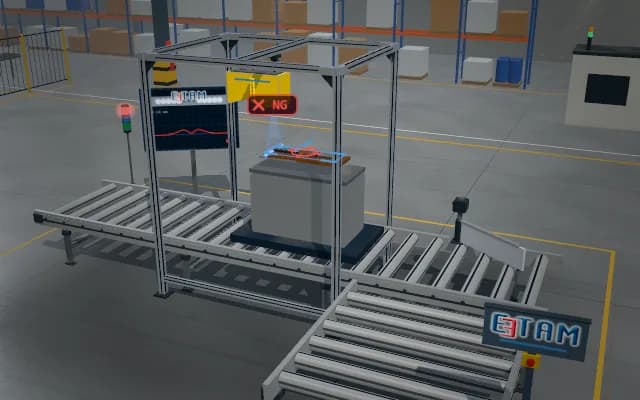
Detecting inactive cells in a cell string
The interconnected cell string is excited inside a light-tight optical tunnel. The emission of the cell regions in the filtered image is compared with one another; a string containing a cell with low optical activity is diverted for rework before lamination.
The position of the suspect cell within the string and its optical activity indicator are recorded. The string is not released to lamination; the vacuum lifting beam moves it to the rework carrier.

- 01 String infeed
- 02 Excite
- 03 Acquire
- 04 Activity
- 05 Route
The string enters the optical tunnel and stops.
- Lowest relative activity
- —
- Suspect cell count
- —
Scenes are illustrative simulations; they are not customer references or performance guarantees.
Need
Brightness differences within a string depend on the distribution of the excitation light as well as on cell activity. Interconnect ribbons and stray ambient light can obscure a low-activity region.
Solution approach
The string receives contactless optical excitation in a light-tight tunnel; a filtered overhead camera collects the photoluminescence image. Cells are split into separate masks and, after correcting for the excitation distribution, their relative emission levels are compared. The result is an optical screening indicator; it does not replace an electrical measurement.
Process steps
- 01
String infeed
The string enters the optical tunnel and stops.
- 02
Excite
Bar lights optically excite the cell string.
- 03
Acquire
The filtered camera collects the emission image of the string.
- 04
Activity
The relative optical response of the cells is compared.
- 05
Route
The string leaves the tunnel; a suspect string is moved to the rework carrier.
Related applications





Let us assess your application together
We test your sample in our laboratory and report on resolution, optics and lighting selection.