
Void analysis in X-ray images of castings
The radiograph received from a separate X-ray system is shown in greyscale. Voids segmented by deep learning are compared with the critical-region mask; E3TAM’s scope is image analysis.
The void mask, critical-region intersection and OK/NG result are displayed; the analysis result goes to the line PLC interface. The cabinet and radiation generation belong to the separate system.

- 01 Sample
- 02 Radiograph
- 03 Mask
- 04 Decide
The casting is positioned on the manipulator and the cabinet door closes.
- Void count
- —
- Critical intersection
- —
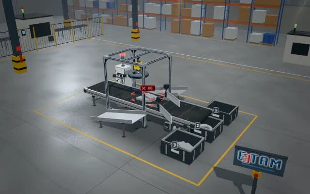
Scenes are illustrative simulations; they are not customer references or performance guarantees.
Need
Changing casting-wall thickness varies the radiograph background. Small gas pores, shrinkage regions and inclusions can blend into these greyscale transitions.
Solution approach
The radiograph of a casting held in the separate X-ray cabinet reaches the analysis display. After background correction, deep learning produces a void mask; its intersection with the critical region is shown over the greyscale image.
Process steps
- 01
Sample
The casting is positioned on the manipulator and the cabinet door closes.
- 02
Radiograph
The separate system’s greyscale image reaches the analysis display.
- 03
Mask
Voids are segmented and intersected with the critical region.
- 04
Decide
The result appears on the display and PLC interface.
Related applications



Let us assess your application together
We test your sample in our laboratory and report on resolution, optics and lighting selection.